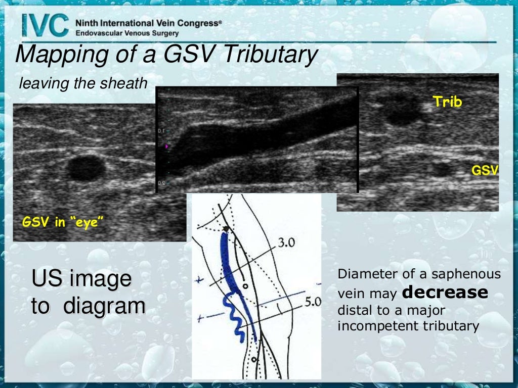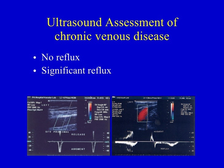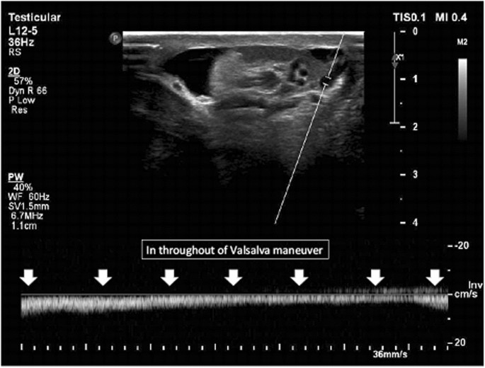

The venous system has two important functions-return of blood to the heart from the capillary bed - maintenance of cardiovascular hemostasis through changes in capacitance. Venous blood pressure is determined by several factors.ĺmong these are pressure generated by the heart, energy lost in the peripheral resistance of arterioles, hydrostatic gravitational forces, blood volume, anatomical composition of the venous wall, effciency of one-way valves, vein wall distensibility (determined by hormonal, systemic alcohol and other�factors), and contraction of venous smooth muscle as influenced by temperature and sympathetic and parasympathetic nerve tone. The International Interdisciplinary Consensus Committee on Venous Anatomical Terminology recommends classifying PVs into six groups according to the segment of the lower extremity in which they are found. 1, Inferior epigastric vein 2, superficial circumflex iliac vein 3, lateral accessory saphenous vein 4, deep external pudendal vein 5, superficial external pudendal vein 6, medial accessory saphenous vein. located 4cm inferior & lateral to pubic tubercle – site of great saphenous vein passing through cribriform fascia (saphenous opening) to reach femoral vein. The most important of superfcial veins are the GSV and the SSV In this slide The great saphenous vein,the longest vein �which courses along the medial aspect of the calf and thigh to join the common femoral vein at the level of the inguinal ligament. According to traditional description, superfcial veins are�separated from deep veins by muscular fascia �These are linked by a variable number of perforator veins which carry blood from the superficial to the deep systems. Normal Flow : Superficial veins drain into the deep veins From theįoot up to the heart Superficial vein disease always starts withĪbnormal valves and interruption to normal flow called venous. #VENOUS REFLUX ULTRASOUND FREE#
Release of proteolytic enzymes and free radicalsĮndothelial damage, tissue destruction, local ischemia Venous hypertension expression of leucocyte adhesion moleculesĪdhesion of WBC to capillary endothelial cells.Raised venous pressure reduced capillary perfusion trapping of WBC.Studies by Raju and Fredericks have shown that this effectĮxplains and correlates with most venous ulceration cases.It contends that reflux is mainly transmitted to the.This theory is the most widespread pathogenesis of CVI.Pigmentation,dermatitis and lipodermatosclerosisĬapillary endothelial damage lack of exchange of nutrients Microlymphatics and dysfunction of local nerve fibersĭefective micro circulation Excessive RBC lysis eczemaĮxcessive release of hemosiderin and fibrin The accumulation of fluid, macromolecules, & extravasated red blood cells

Microcirculatory dysfunction in chronicĬapillaries of patients with CVI increased pericapillary edema
Failure of valves located at junctions ofĬhronic damage and microcirculatory dysfunction. Dysfunction or incompetence of the valves. Karthik Gujja, MD, MPH, Jose Wiley, MD*, Prakash Krishnan, MD : Chronic Venous Insufficiency :Ĭlinic review article, Peripheral vascular disease Rev 2014 Refill occurs rapidly by both arterial inflow and Preexisting weakness in the vessel wall orĮxcessive venous distention resulting fromīlood volume is pumped out of the extremity, These mechanisms induce venous hypertension, Venous pressure is increased and return of blood is. VEINS DISTEND,ELONGATE,TORTOUS,POUCHED,INELASTIC STRECHING OF VALVES-VALVULAR INCOMPETENCE ANY RISK FACTOR INCREASED VENOUS PRESSURE. Anatomy of the veins of the lower extremity. Chronic venous insufficiency have two component. Medical condition where veins cannot pump enough. Chronic venous insufficiency -DEFINITION. Concept of Venous Microcirculatory Disturbances and. Concept of Macrocirculatory Dysfunction. Basic concept of venous anatomy and physiology of venous. Jünger M1, Steins A, Hahn M, Häfner HM. Insufficiency : clinic review article, Peripheral vascular disease Rev 2014 
Karthik Gujja, MD, MPH, Jose Wiley, MD*, Prakash Krishnan, MD : Chronic Venous.Quantifying saphenous reflux : Journal of vascular surgery venous and lymphaticĭisorder, The American venous forum 2015

Seshadri Raju, MD, FACS,a Mark Ward Jr, MS,a and Tamekia L. Pathophysiological basis and clinical perspectives. Chiu J.J., and Chien S.: Effects of disturbed flow on vascular endothelium:.


 0 kommentar(er)
